Leg Bone Diagram - Infographic Diagram Of Human Femur Bone Or Leg Bone ... : Our goal is that these leg anatomy worksheets pictures gallery can be a direction for you, bring you more references and also make you have a great day.. Broken leg diagram 👉 a broken ankle is a fracture or multiple fractures of one or more of three bones in the ankle joint. Includes leg (femur, tibia, patella, and fibula) and foot (tarsals and digits) bones. Ankle & lower leg anatomy. The z type of leg joint does not need the scale bone to control its movement. Leg femur diagram data wiring diagram today.
Image result for leg bones diagram human leg bone structure your leg bones are the longest and strongest bones in your body. The foot bones shown in this diagram are the talus, navicular, cuneiform. Distal to the ankle is the foot. Master leg and knee anatomy using our topic page. The knee joint is the largest joint in the body and is primarily a hinge joint, although some sliding and rotation occur.
Blank Leg Bone Diagram - 19 1 Types Of Skeletal Systems ... from st3.depositphotos.com The tibia and the fibula, at the top of the ankle joint. The femur, or thighbone, is the longest and largest bone in the human body. Leg bones diagram diagram schematic ideas lower leg muscle diagram blank sketch coloring page antique 1890s medical anatomy diagram leg bones skeleton posted on april 18, 2019april 18, 2019. The femur, or thighbone, is the longest and largest bone within the human physique. Our goal is that these leg anatomy worksheets pictures gallery can be a direction for you, bring you more references and also make you have a great day. The smaller lateral bone of the lower leg. Your leg bones are the longest and strongest bones in your body. The tibia, commonly known as the 'shin bone', is the largest and most medial of the two.
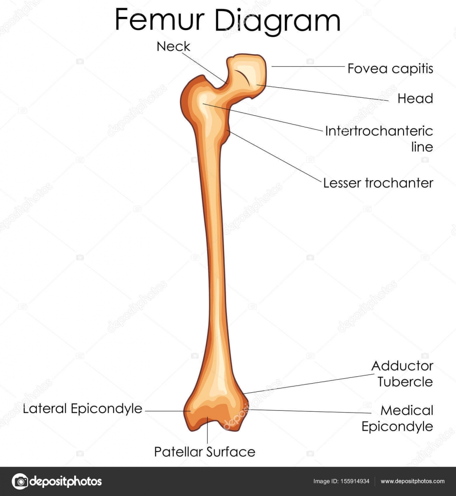
The femur, or thighbone, is the longest and largest bone in the human body.
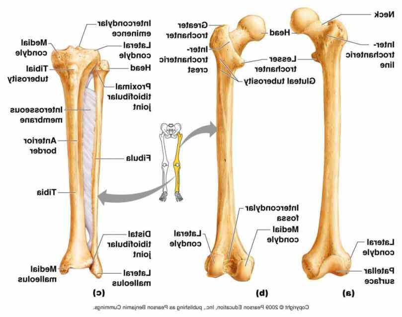
Includes leg (femur, tibia, patella, and fibula) and foot (tarsals and digits) bones. The z type of leg joint does not need the scale bone to control its movement. Start studying leg bone labeling. The majority of muscles in the leg are considered long muscles, in that they stretch great distances. Leg bones diagram diagram schematic ideas lower leg muscle diagram blank sketch coloring page antique 1890s medical anatomy diagram leg bones skeleton posted on april 18, 2019april 18, 2019. The hip itself is a ball and socket joint, much like the shoulder.the structures necessary to create this joint are the socket, the joint capsule, muscle, ligaments, and the neck. Frog leg bones diagram / comparative vertebrate anatomy lecture notes 5 : Bone diagram forehead (frontal bone) nose bones (nasals) cheek bone (zygoma) upper jaw (maxilla) lower jaw (mandible) breast bone (sternum) upper arm bone (humerus) lower arm bone (ulna) thigh bone (femur) collar bone (clavicle) toe bones (phalanges) ankle bones (tarsals) kneecap (patella) shin bone This diagram depicts diagram leg bones anatomy.human anatomy diagrams show internal organs, cells, systems, conditions, symptoms and sickness information and/or tips for healthy living. You can palpate its anterior border when you run your finger down the anterior aspect of your leg. The bones of the leg are the femur, tibia, fibula and patella.the foot bones shown in this diagram are the talus, navicular, cuneiform, cuboid, metatarsals and calcaneus. It is the largest bone in the body and is the only bone in the upper leg. The thigh bone, or femur, is the large upper leg bone that connects the lower leg bones (knee joint) to the pelvic bone (hip joint).
Human leg bone diagram : The thigh bone, or femur, is the large upper leg bone that connects the lower leg bones (knee joint) to the pelvic bone (hip joint). Cross section of foot nerves 13 photos of the cross section of foot nerves cross section of nerve fiber, foot anatomy nerves, foot nerve pain, human foot nerves, nerve cross section histology, peripheral nerve cross section, spinal nerve cross section, foot, cross section of nerve fiber, foot anatomy nerves, foot. Ankle & lower leg anatomy. They allow you to move and provide support for your upper body.
Leg Bones - Medical Art Library from www.medicalartlibrary.com Some types of leg pain can be traced to problems in your lower spine. The lower leg extends from the knee to the ankle. Pin on medical websites we like. Distal to the ankle is the foot. These are the femur, patella, tibia, fibula, tarsal bones, metatarsal bones, and phalanges (see figure 6.51). Its decrease finish helps create the knee joint. Leg femur diagram data wiring diagram today. The hip joint gives the leg an incredible range of motion while still providing support to the body's weight.
Examine the hind legs and front legs of the frog.
Browse 7,069 leg bone stock photos and images available, or search for human leg bone or leg bone xray to find more great stock photos and pictures. Beside that, we also come with more related ideas as follows free printable human anatomy coloring pages, lower leg muscle diagram blank and lower limb bones unlabeled. They allow you to move and provide support for your upper body. The bones together make up the hip. The foot bones shown in this diagram are the talus, navicular, cuneiform. Image result for leg bones diagram human leg bone structure your leg bones are the longest and strongest bones in your body. The tibia and the fibula, at the top of the ankle joint. The femur is known as a long bone. It is the largest bone in the body and is the only bone in the upper leg. Bone diagram forehead (frontal bone) nose bones (nasals) cheek bone (zygoma) upper jaw (maxilla) lower jaw (mandible) breast bone (sternum) upper arm bone (humerus) lower arm bone (ulna) thigh bone (femur) collar bone (clavicle) toe bones (phalanges) ankle bones (tarsals) kneecap (patella) shin bone The smaller lateral bone of the lower leg. (there are four types of bone: Master leg and knee anatomy using our topic page.
Distal end of right humerus. At the distal end of the femur, two rounded condyles meet the tibia and fibula bones of the lower leg to form the knee joint. The medial, larger bone of the lower leg. Beside that, we also come with more related ideas as follows free printable human anatomy coloring pages, lower leg muscle diagram blank and lower limb bones unlabeled. The femur is the single bone of the thigh.
Anatomy The Bones Of The Lower Limb | MedicineBTG.com from medicinebtg.com The femur, or thighbone, is the longest and largest bone within the human physique. This area is commonly referred to as the calf. At the distal end of the femur, two rounded condyles meet the tibia and fibula bones of the lower leg to form the knee joint. Ankle & lower leg anatomy. The pubis, ischium, and ilium together constitute the pelvis while the thigh bone is the femur. Related posts of leg bones anatomy diagram cross section of foot nerves. (there are four types of bone: Its lower end helps create the knee joint.
These are the femur, patella, tibia, fibula, tarsal bones, metatarsal bones, and phalanges (see figure 6.51).
The bones of the hip include the femur, the ilium, the ischium, and the pubis. The tibia and the fibula, at the top of the ankle joint. It is the largest bone in the body and is the only bone in the upper leg. Most leg pain results from wear and tear, overuse, or injuries in joints or bones or in muscles, ligaments, tendons or other soft tissues. The bones of the leg are the femur, tibia, fibula and patella.the foot bones shown in this diagram are the talus, navicular, cuneiform, cuboid, metatarsals and calcaneus. The femur, or thighbone, is the longest and largest bone in the human body. Master leg and knee anatomy using our topic page. Includes leg (femur, tibia, patella, and fibula) and foot (tarsals and digits) bones. Image result for leg bones diagram human leg bone structure your leg bones are the longest and strongest bones in your body. Related posts of diagram of leg bones inside of arm muscle and bone. Inside of arm muscle and bone 12 photos of the inside of arm muscle and bone , bone Long bones, short bones, flat bones, and irregular bones.) long bones are longer than they are wide, with spongy bones at both ends and a cavity filled with bone marrow in the shaft. The tibia and fibula are two long bones that run parallel to each other, forming the scaffold of the leg and providing attachment points for many muscles.